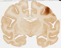
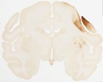
Datasets -> Macaca Mulatta

[ Macaca Mulatta General Info ]

RM311: V1 inj.

CTb tracer injection made into foveal representation of V1 of left hemisphere. There are two microinjection tracks approximately 2mm apart seen in sections 132-142 and 145-158. Both tracks enter the white matter beneath area 17.

The cortical injection is confined to area 17 as defined by comparison with the adjacent Nissl-stained sections. (The anterior border of area 17 ends in section 163). The injection is heaviest in sections 115-146.

Anterograde labeling commences in area 18 in section 142. Retrograde labeling of cells in layers III and VI of area 18 commences in section 154. Labeling in area 18 ends in section 183.

Retrograde labeling of neurons commences in area MT in section 165 and anterograde labeling in section 193.

Some labeling can be seen in area V3/4 in sections 195-225 and a few retrogradely labeled layer VI cells can be seen in the floor of the superior temporal sulcus in sections 230-243.

There are three spots of anterograde and retrograde labeling in the medial and lateral divisions of the inferior pulvinar nucleus in sections 234-239, and a single patch in the dorsal lateral geniculate nucleus in sections 244-248.

RM311: V1 inj.
coronal, whole-brain

507 slides, 0.46um/pixel
Dataset Id: 121
[ Metadata ]
RM311: Nissl
coronal, whole-brain

506 slides, 0.46um/pixel
Dataset Id: 122
[ Metadata ]
RM312: MI inj.

RM312 MI injection

The injection of CTB is centered in the middle of cytoarchitectonic area 4 at about the middle of its medio-lateral extent and immediately posterior to the arcuate sulcus. The maximal extent of the injection can be seen on sections 156-166. Anteriorly, there may be some encroachment on area 6 in the posterior bank of the arcuate sulcus.

Going anteriorly from the injection (diminishing section numbers from section 145), cellular labeling in area 4 itself is quite localized. There are distinct patches of coincident anterograde and retrograde labeling in area 6 below and posterior to the arcuate sulcus and in medial area 6 above the cingulate sulcus and adjacent to the area 4 border. There are a few scattered labeled cells in the cingulate gyrus (area 24).

Over the course of the injection and continuing posteriorly (sections 139-189), there is a distinct patch of retrograde and anterograde labeling along the fundus of the cingulate sulcus in what is probably a further division of area 6.

Posterior to the injection (increasing section numbers from section 170), there are two patches of anterograde and retrograde label in area 4, with a few scattered labeled cells elsewhere in area 4, their numbers declining posteriorly (sections 205-218). Retrograde labeling of cells in the upper layers of areas 1 and especially 2 can be seen in sections 220-250. There are also patches of labeled cells in the medial bank of the intraparietal sulcus (area 2 or 5) and in the upper bank of the lateral sulcus (area SII). Further posteriorly (sections 259-269) there is a distinct patch of coincident retrograde and anterograde labeling in area 7 in the medial bank of the intraparietal sulcus. This may reflect involvement of part of area 6 in the injection.

Elsewhere, there is retrograde labeling of cells throughout the greater part of the claustrum (sections 170-228), a patch of anterograde label in the putamen in sections 180-201, and in the thalamus patches of retrograde and anterograde labeling can be seen in the VLp nucleus in sections 222-245 and in the CL nucleus in sections 240-255.

RM312: MI inj.
coronal, whole-brain

200 slides, 0.46um/pixel
Dataset Id: 154
[ Metadata ]
RM312: Nissl
coronal, whole-brain

473 slides, 0.46um/pixel
Dataset Id: 155
[ Metadata ]
RM315: area 7 inj.

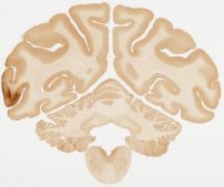
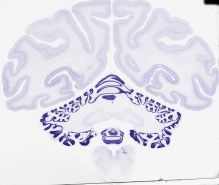
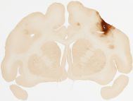
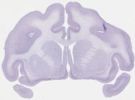
Primary CTb tracer injection made into upper bank of intraparietal sulcus (area 7) in the left hemisphere (sections 218-232). A smaller and more ventrally located injection is restricted to deeper layers of area 7 (with some white matter included) (sections 236-239).

Anterograde label can be seen in cortex, thalamus, and basal ganglia.

In cortex, anterograde label is seen in inferior occipital gyrus (sections 236-243) and inferior postcentral gyrus (SII) (sections 164-175).

In thalamus, dense anterograde label in centrolateral nucleus (CL) and the anterior (Pla) and medial (Plm) nuclei of the pulvinar begins in section 246 and ends in section 262.

In basal ganglia, patchy anterograde label is seen in sections 221-241.

Retrograde labeled cells were seen in multiple ipsilateral cortical and subcortical areas.

In cortex, labeled cells were seen in inferior postcentral gyrus (sections 190-229), cingulate gyrus (sections 150-231), occipital temporal gyrus and inferior occipital gyrus (sections 245-266), and inferior temporal gyrus (sections 201-209 and 226-244). A group of labeled cells was seen within the anterior portion of secondary somatosensory cortex (bordering the subcentral sulcus) in continuation to the inferior postcentral gyrus (sections 164-175). Labeled cells were seen in insular cortex (sections 173-196).

Labeled cells are seen in multiple thalamic nuclei. Some cells were seen in ventromedial region of lateral dorsal (LD) nucleus (sections 215-219). Some cells of the intralaminar nuclei were labeled in rhomboid (Rh) and central medial (CeM) (sections 206-213), central lateral (CL) (sections 218-239) and paracentral (sections 225-233) nuclei. Within pulvinar labeled cells were seen in the anterior (Pla) and medial (Plm) nuclei (sections 246-262). Labeled cells were also seen in field of forel (FF) (sections 219-227).

Other subcortical labeled cells are seen in claustrum (sections 175-240) and basal forebrain (sections 137-159).

Retrograde labeled cells were seen in contralateral cortex. Labeled cells were located in contralateral area 7 in upper layers of the anterior marginal gyrus (AMG) (sections 201-254). Labeled cells were also seen in contralateral cingulate cortex (sections 145-153).

RM315: area 7 inj.
coronal, whole-brain

267 slides, 0.46um/pixel
Dataset Id: 124
[ Metadata ]
RM315: Nissl
coronal, whole-brain

452 slides, 0.46um/pixel
Dataset Id: 153
[ Metadata ]
RM316: SI inj.

RM316: SI inj.
coronal, whole-brain

462 slides, 0.46um/pixel
Dataset Id: 158
[ Metadata ]
RM316: Nissl
coronal, whole-brain

466 slides, 0.46um/pixel
Dataset Id: 159
[ Metadata ]